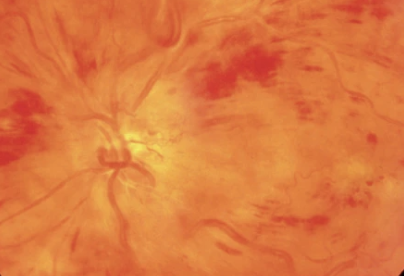
Central retinal vein occlusion (CRVO)
- Ischemic (20–35%): absent capillary perfusion/ischemia > 10 disc diameters
- Non-ischemic: < 10 disc diameters
History
- Decreased visual acuity, visual field defects, metamorphopsia
- Risk factors: arterial hypertension 30–60%, coronary artery disease 22–50%, dyslipidemia 30–60%, diabetes mellitus 14–34%, glaucoma, smoking, obesity, contraceptives
- Further risk factors: syphilis, sarcoidosis, vasculitis, hyperviscosity syndromes (multiple myeloma, leukemia, Waldenström's macroglobulinemia), sickle cell disease, HIV, hyperhomocysteinemia, elevated ESR
Examinations
- RAPD
- Visual field, finger perimetry
- Subjective visual acuity
- Applanation tonometry and optic disc assessment
- Anterior segment (iris rubeosis)
- Fundus in mydriasis (macular edema, flame-/blot-shaped hemorrhages, exudates, dilated/tortuous veins, disc swelling), OCT (macular edema, disc swelling)
Work-up
Patient > 50 years
> Work-up for cardiovascular risk factors, rule out glaucoma
- Intraocular pressure: diurnal pressure curve
- Blood pressure
- Blood tests: CRP, differential blood count, ESR (> typically elevated in women), hematocrit (elevated blood viscosity), prothrombin time, glucose, HbA1c, fasting lipid panel
- Smoking status
Patient < 50 years
> Same work-up as for patients > 50 years, but additionally:
- Differential blood count (polycythemia, essential thrombocythemia, leukemias, hemoglobinopathies)
- Serum protein electrophoresis (> paraproteinemias, e.g. Waldenström's macroglobulinemia)
- Coagulation work-up (via hematology, only after 2 months, in the absence of risk factors or in multiple/bilateral CRVO): anticardiolipin antibodies, antiphospholipid antibodies, activated protein C resistance, homocysteine, antithrombin III, protein S, protein C)
- ANA, rheumatoid factor (> rheumatic and non-rheumatic diseases)
- ACE (> sarcoidosis)
- Treponema pallidum (> syphilis, "syphilis screening test")
Clinical course (including screening for neovascularization)
- Fluorescein angiography after resorption of extensive hemorrhages (usually after 3 months in the retina clinic) to rule out areas of non-perfusion
- Photo documentation (posterior pole, before laser therapy)
- Internal medicine work-up by the primary care physician to rule out/optimize cardiovascular risk factors
Treatment
Acute phase (up to 8 weeks)
- Lowering of intraocular pressure (even if only mildly elevated!)
- Aspirin 100mg tablet 1x/day (evidence not established)
- Intravitreal anti-VEGF therapy or intravitreal steroids if needed (for macular edema): see treatment pathway in the IVT chapter
- Treatment of underlying systemic conditions
- Consider replacing diuretics with other antihypertensive medications
- Discontinue any oral contraceptives
- Counsel about isovolemic hemodilution (no evidence)
- Blood cells are removed from the blood via phlebotomy and volume replacement > hematocrit decreases (below 37%) > blood becomes "less viscous"
Chronic phase (complications)
- Areas of non-perfusion, secondary glaucoma > upon development of neovascularization of the iris, angle, disc, or retina
- Fluorescein angiography after resorption of hemorrhages (4–12 weeks)
- Panretinal laser photocoagulation (laser parameters: 500 µm, 0.2 sec, outside the vascular arcades to anterior of the equator, total of 1200–1500 spots over approx. 3 sessions, max. 500 spots per session)
- In macular edema, no improvement in visual acuity is to be expected from focal laser photocoagulation
- Consider anti-VEGF: see treatment pathway in the IVT chapter
- Consider cyclophotocoagulation
Follow-up
- Ischemic CRVO: monthly for the first 6 months (caution: approx. 60% develop neovascularization!)
- Non-ischemic CRVO: monthly for the first 6 months (caution: approx. 1/3 convert to ischemic CRVO within the first year!)
Visual prognosis
- Ischemic CRVO: over 90% have visual acuity of 0.1 or worse, neovascular glaucoma in over 60% of cases within weeks to years
- Non-ischemic CRVO: 10% of cases show uncomplicated resolution of the occlusion, 50% have visual acuity of 0.1 or worse, approx. 1/3 progress to ischemic CRVO within one year
Branch retinal vein occlusion (BRVO)
Work-up > as for CRVO
Treatment
- Treatment of underlying systemic conditions
- In macular edema, anti-VEGF therapy is indicated
- After resolution of the hemorrhages, fluorescein angiography should be performed.
Follow-up
- Follow-up examinations are performed monthly for the first 6 months. Then every 3 months up to one year after the event.
Visual prognosis
53% achieve 0.5 or better, 25% between 0.2 and 0.4, 22% 0.1 or worse.
> Patient information
Source: Will's Eye Manual, Eyewiki AAO