History
- Acute vision loss (naAION: painless), visual field defects (horizontal), initially unilateral, bilateral involvement possible later, > 55 years,
- possibly CRAO/cranial nerve palsy (abducens palsy)
- Cardiovascular risk factors, hyperlipidemia, arterial hypertension, obstructive sleep apnea syndrome
- Arteritic AION (aAION): jaw claudication, scalp tenderness on combing, headache, arthralgias, weight loss, B symptoms, poor general condition
Differential Diagnosis
Central or hemi-central retinal artery occlusion (no disc swelling), central retinal vein occlusion (retinal hemorrhages extending to the periphery), optic neuritis (hyperemic disc edema),
Diagnostics
- RAPD (positive)
- Ishihara color vision testing, red desaturation
- Subjective visual acuity
- Applanation tonometry
- Anterior segment (aAION: thickened, tender temporal artery, tender scalp)
- Fundus examination under mydriasis (retinal changes, disc assessment
- OCT of the macula and optic disc and photographs (to exclude differential diagnoses)
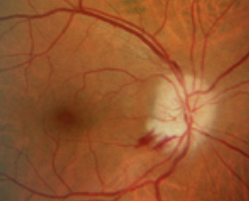
- Disc edema (acute), optic atrophy without increased cupping (older optic neuropathy)
- Blood tests (note: if aAION is suspected > emergency referral to a tertiary center for further work-up and treatment if indicated)
- Erythrocyte sedimentation rate (ESR): men (age/2), women (age+10/2) > normal in 10% despite aAION
- CRP (age-independent)
- Complete blood count, AST/ALT, ALP, GGT, LDH, creatinine, glucose, INR
If aAION is Suspected
- Neurology: neurovascular ultrasound: assess for flow reversal (before temporal artery biopsy
- Internal medicine: assess for contraindications to high-dose steroid therapy
- Temporal artery biopsy if giant cell arteritis is suspected, within one week of starting steroid therapy)
- If the biopsy is negative but suspicion remains high > biopsy of the contralateral side
If naAION is Suspected
- Rheumatologic work-up: assess for vasculitis, vascular ultrasound?
- Additional blood tests per rheumatology
Prognosis
aAION
- without treatment: 54-95% vision loss within 4 months, up to 50% in the fellow eye
- with steroid therapy: 13% vision loss and prevention of vision loss in the fellow eye
Treatment
aAION
- Inpatient admission to a tertiary center: intravenous methylprednisolone for 3 days (in the morning), then switch to oral prednisone 60-100mg/day
- PPI (esomeprazole), Calcimagon
- after discharge: gradual tapering of steroid therapy guided by CRP/ESR, managed by the primary care physician or rheumatologist (over several months)
- switch to steroid-sparing therapy if applicable (e.g. methotrexate)
naAION
- Aspirin 100mg tablet 1x/day (no evidence for a positive long-term effect)
- Primary care physician: evaluate cardiovascular risk factors, diabetes mellitus, arterial hypertension, sleep apnea, consider taking antihypertensives in the morning instead of the evening if applicable (nocturnal hypotension)
Follow-up
naAION: after one month with subjective visual acuity, OCT of the macula and optic disc and photographs, visual field (driving fitness assessment)
aAION: after discharge from the tertiary center, close ophthalmologic follow-up with blood tests (CRP/ESR) by the primary care physician/rheumatologist
Sources: The Will's Eye Manual, Eyewiki.org