Informed consent -> see consent form for pterygium excision (incl. infection/scarring/postoperative pain and inability to work for at least 1-2, up to 4 weeks)
Preoperative preparation
Oxybuprocaine 0.4% eye drops 3x every 5 minutes
Surgical Materials
Secure the UV device firmly to the edge of an instrument table with a clamp, plug in the cable safely, calibrate if necessary
Pachymeter: insert batteries and switch on
Hockey knife
Disposable Materials
Hibidil® solution
Round swabs x6
Sterile drape
Disposable film scissors
Bangerter lid speculum
BSS solution in a 10 ml syringe with a blunt cannula
Sterile distilled water in a 10 ml syringe with a blunt irrigation cannula
Lidocaine 2% solution
Riboflavin 0.1% HPMC solution (e.g. VibeX Rapid®)
Triangular swabs
Bandage contact lens
Soaked in Dexafree eye drops 2x and Floxal eye drops 2x for at least 30 minutes
Floxal eye ointment
Surgical Procedure
Topical anesthesia. Disinfection. Sterile draping. Corneal abrasio using the hockey knife. +PLUS (self-pay service): topography-/aberrometry-guided PTK/transPTK. Ultrasound pachymetry. Preparation of the stroma for 20 minutes by alternating application of riboflavin solution and distilled water every two minutes. Oxybuprocaine eye drops every five minutes. Irradiation with UVA according to the respective protocol +PLUS (self-pay service): customized protocol Rinsing of the cornea with BSS solution. Floxal eye ointment applied to the cornea, 2 mm ointment strand. Insertion of the loaded bandage contact lens. Application of a monocular patch with light pressure.
caution:
working distance from the UVA source to the corneal apex = 50 mm -> the irradiated area is sharply demarcated
the irradiated area should be centered over the pupil center, or slightly decentered according to the location of the keratoconus (usually nasal-inferior); do not irradiate the limbus
the patch must be applied with light pressure so that the patient cannot blink under the patch
Surgical Protocols
Standard at ONO: Accelerated Protocol
Riboflavin application 20 min,
Intensity 9 mW/cm²
Treatment duration 10 min
PRK+CXL
Accelerated Protocol (see above)
Topography-guided PRK
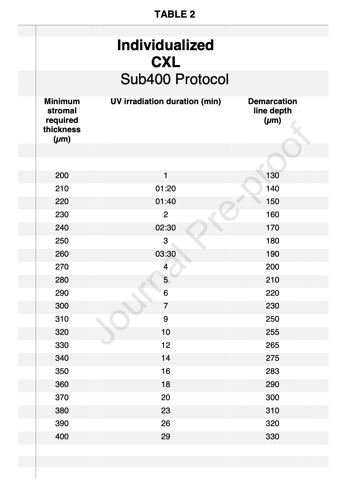
sub400 Protocol (pachymetry 200-400 µm)
Treatment duration is adjusted to corneal thickness
Settings
Intensity 3 mW/cm²
Epi-off
Chromophore 0.1% riboflavin, swelled with distilled water
Irradiation mode: continuous
UV source: CXL-365
Treatment time depends on pachymetry
Individual fluence, 10 µm steps
Postoperative
Analgesia (caution: adjust if necessary for body weight and liver/kidney values)
Paracetamol 500 mg tablet 4x/day up to 8x/day (e.g. Dafalgan®)
Non-steroidal anti-inflammatory drug (e.g. mefenamic acid 500 mg tablet 3x/day or ibuprofen 400 mg tablet 4x/day)
4 days: remove contact lens if applicable (caution: carefully, from inferior with a cotton-tipped applicator or with fine anatomical forceps from the temporal side, patient looks nasally)
Abrasion re-epithelialized: start with FML eye drops 4x/day for 1 week, 3x/day for 1 week, 2x/day for 1 week, 1x/day for 1 week
Persistent erosion: follow-up every two days until complete epithelial closure, only then start FML eye drops
Showering possible again, no water or shampoo in the eye, do not rub, eye shield at night
2 weeks
Corneal OCT central volume scan (measuring the depth of the demarcation line, given as a percentage of total corneal thickness, e.g. 80%)
No sauna/steam bath/swimming pool/diving underwater
1 month
Topography, corneal OCT (only for documentation if haze/scarring present)
Subjective visual acuity
If haze present, increase FML eye drops again to 4x/day for 2 weeks -> follow-up with corneal OCT
taper over 8 weeks (3x/day for 2 weeks, etc.)
All activities possible again
if applicable, clear patient for fitting of rigid gas-permeable contact lenses (prescription with MiGel position 25.02.03.00.1)
4 months
Topography, corneal OCT (only for documentation if haze/scarring present)
Subjective visual acuity
If late-onset haze present, increase FML eye drops again to 4x/day for 2 weeks -> follow-up with corneal OCT, taper over 8 weeks (3x/day for 2 weeks, etc.)
If the cornea has cleared and there is existing progression of ectasia in the fellow eye -> CXL on the fellow eye
FAQs
Should CXL be performed early in young patients with early keratoconus?
Patients aged 14-25 years -> ectasia (keratoconus) can progress rapidly
In patients who frequently rub their eyes -> start antiallergic medication
Opatanol eye drops 2x/day and Telfast 120 mg tablet 1x/day
Follow-up with topography after 3 months
If progression -> case conference -> CXL
Should an eye with progressive ectasia but still very good best-corrected visual acuity be treated with CXL?
Yes, because the refractive error will soon no longer be correctable due to progressive corneal bulging. This can be prevented by stopping the progression.
Do rigid gas-permeable contact lenses help stop the progression of keratoconus?
No, this is a misconception. Keratoconus continues to progress underneath the contact lens.
Contact lens removal before topography
Rigid gas-permeable contact lenses can distort the epithelium and thus the topography -> contact lens removal for 1 week for soft lenses and 2 weeks for rigid lenses. If this is not feasible for the patient because visual acuity cannot be adequately corrected with spectacles:
Separate follow-up visits for each eye with the same lens-free period
Reduce the lens-free period to at least 3 days (e.g. Friday to Monday, follow-up on Monday), if necessary certify inability to work
Can thin corneas be treated?
Yes. Thanks to the sub400 protocol, corneas with pachymetry down to 200 µm can be treated. For this, the intensity is reduced to 3 mW/cm² (as in the Dresden protocol) and, for example at 200 µm (thinnest location), irradiation is applied for only one minute (see above).
When does CXL no longer make sense?
As soon as dense stromal opacities appear and visual acuity can no longer be corrected to above 0.5 even with a pinhole or rigid gas-permeable contact lenses. In these cases, vision can only be rehabilitated by keratoplasty.
You might also be interested in this
Cornea
conjunctival cyst
Conjunctival cysts must be completely removed to minimize the risk of them recurring
Cornea
Pterygium and treatment
The conjunctiva grows over the cornea and can threaten vision
Cornea
Salzmann Knots
Salzmann's nodules on the cornea should be removed early