9.10.2024
Reading time:
10 Min.

Diabetic Retinopathy Management

Classification, management

Dr. Valery Vinzent Wittwer

Diabetic Retinopathy (DR)

History

  • Initial diagnosis of diabetes mellitus and treatment
    • Poorly controlled diabetes: target HbA1c approx. 7%
  • Arterial hypertension: rigorous blood pressure control recommended
  • Renal insufficiency: ask about renal function values
  • Dyslipidemia
  • Pregnancy

Differential diagnosis

Central retinal vein occlusion, branch retinal vein occlusion, ocular ischemic syndrome, hypertensive retinopathy, radiation retinopathy

Classification (ETDRS), management

  • No diabetic retinopathy in a patient with known diabetes mellitus
    • ONO: Annual follow-up with OCT M P and photographs
    • ONO: Gestational diabetes: Follow-up in each trimester with OCT M P and photographs
Non-proliferative diabetic retinopathy
  • Mild non-proliferative diabetic retinopathy (NPDR):
    • Microaneurysms
  • Moderate NPDR, additionally:
    • Retinal hemorrhages (20 per quadrant in 1–3 quadrants)
    • Mild intraretinal microvascular abnormalities (IRMA)
    • Venous beading in > 1 quadrant
    • Cotton-wool spots
    • >  Follow-up in 6 months (26% > PDR, 8% PDR within 12 months)
  • Severe NPDR: presence of one of the following features (= 4-2-1 rule), additionally:
    • Extensive hemorrhages and microaneurysms in 4 quadrants
    • Definite venous beading in at least 2 quadrants
    • Moderate IRMA in at least 1 quadrant
    • > Follow-up in 4 months (50% > PDR, 15% > PDR within 12 months)
    • > Fluorescein angiography (FA), if needed scattered panretinal laser photocoagulation
  • Very severe NPDR
    • 2 criteria of severe NPDR
    • > Follow-up in 2–3 months (45% > PDR within 6 months)
Proliferative diabetic retinopathy (PDR):
  • Mild to moderate PDR
    • Neovascularization (of the disc / NVD = Optic Disc
    • > Follow-up in 2 months
    • > Scattered panretinal laser photocoagulation
  • High-risk group
    • NVD (>1/3 disc area)
    • NVD (any) + preretinal or vitreous hemorrhage
    • NVE (> 1 disc diameter (DD)) + preretinal or vitreous hemorrhage
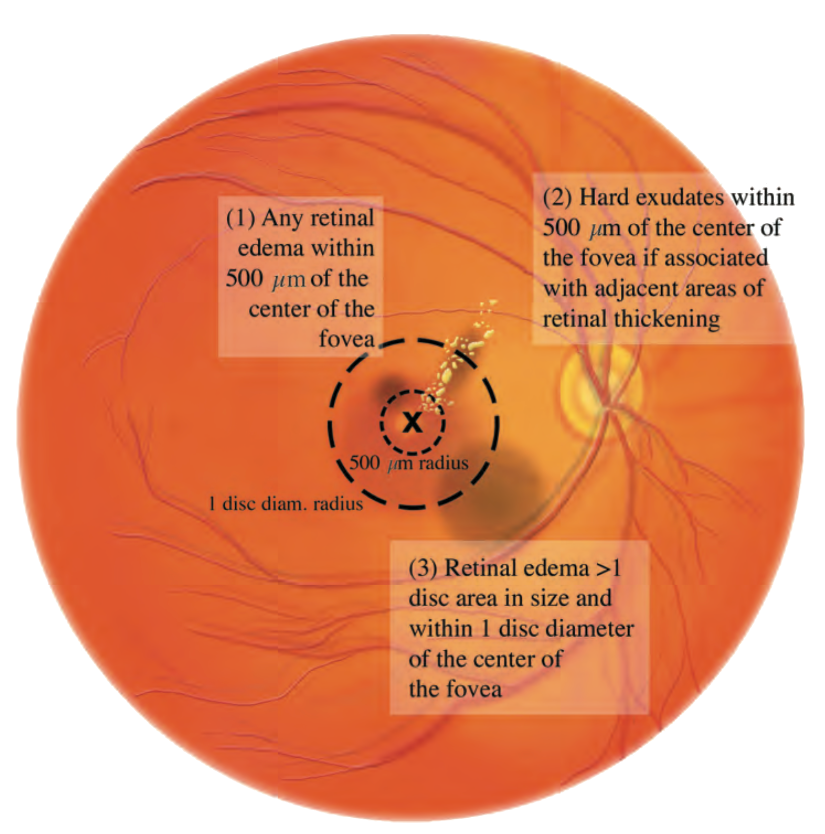
  • With clinically significant macular edema (ETDRS):
    • Edema or hard exudates within 500 μm of the fovea
    • Retinal thickening > 1 DD within 1 DD of the fovea
    • Decreased visual acuity due to edema
    • > IVT with anti-VEGF following a treat-and-extend regimen
__wf_reserved_inherit
Indication for IVT in diabetic macular edema
1. Macular edema within 500 μm of the fovea
2. Hard exudates and retinal thickening within 500 μm of the fovea
3. Retinal edema greater than 1 disc diameter in size located within 1 disc diameter of the fovea

Intravitreal therapy

Low responder: insufficient response to anti-VEGF after 6–12 months

  • Switch to Ozurdex
  • Absolute contraindication (CI):
    • Known steroid response
  • Relative CI: 
    • Iridotomy (possible migration of the Ozurdex implant into the anterior chamber > endothelial cell loss > counsel the patient accordingly and sensitize them to symptoms
    • Phakic patient (> cataract)

In case of vitreous hemorrhage: evaluate pars plana vitrectomy (PPV)

Sources: Will's Eye Manual, Eyewiki.org, Blaubuch Inselspital

You might also be interested in this

Retina

venous occlusion

Eye thrombosis can lead to rapid loss of vision and later also to glaucoma with painful blindness. Early diagnosis and treatment can significantly improve the course of this disease and preserve vision.
Retina

Arterial occlusion

Painless blindness. The occlusion of a retinal artery is comparable to a stroke. Prompt treatment can save vision and prevent further episodes.