Cataract surgery removes the natural crystalline lens and implants an artificial lens (IOL), both in a single operation. The refractive power of the IOL therefore has to be determined in advance. The following parameters are used to determine IOL power:
Axial length (AL) in millimetres
Keratometry values (K) in dioptres
Anterior chamber depth (ACD) in millimetres
For a more precise determination, newer formulas (fourth and fifth generation) also predict the anticipated IOL position in the eye (Effective Lens Position) using ray-tracing, artificial intelligence, or regression
Target refraction
Distance (emmetropia): focal point at distance (> 6 metres to infinity) for watching TV or driving without glasses -> 0 dioptres
Intermediate distance (slight myopia) for computer distance and reading the newspaper on the table, or for monovision: focal point at intermediate distance -> -1.5 dioptres (66 centimetres)
Reading distance (myopia) for reading books -> -2.0 (50 centimetres) to -2.5 dioptres (40 centimetres) to -3.0 dioptres (33 centimetres)
Monovision
Determine the dominant eye
Simulate monovision with a trial frame ("plug-in glasses")
1. Fully correct both eyes for distance, target refraction 0 dioptres
2. Correct the non-dominant eye for near with +1.5 (convex plus lens) -> target refraction -1.5 dioptres
3. Simulate the opposite (dominant eye for near)
4. Have the patient look into the distance (visual acuity chart) and at near (reading chart) with the trial frame
5. Send the patient to the waiting room with the trial frame and instruct them to look around and walk about with it to get a spatial impression of monovision
6. Evaluate the patient's feedback
Positive feedback: it works well, initially still unfamiliar but relatively comfortable after a few minutes -> surgery can be planned
Positive feedback, but: the patient is very uncertain -> contact lens trial
Unclear feedback: it works but feels very unfamiliar -> contact lens trial or multifocal IOLs
Negative feedback: neither distance nor near vision is sharp, has to close one eye to see clearly with the other, cannot judge distances -> multifocal IOLs
Monovision with anisometropia of more than 1.5 dioptres
Reduced three-dimensional perception
In principle only in cases of:
Pre-existing anisometropia
Amblyopia (despite the amblyopia, the amblyopic eye sees better at near than the better eye -> stimulation -> possible slight improvement in visual acuity)
Correcting astigmatism in monovision
Undercorrect the near eye (leave 0.5 dioptres of astigmatism) -> improved depth of focus
Caution: new anisometropia can make binocular fusion more difficult and thereby aggravate a latent squint (phoria) -> test with a trial frame, contact lens trial if necessary
Simulation with contact lenses
If the patient wants monovision but is unsure, a contact lens wearing trial should be carried out
From 1 dioptre of astigmatism onward, this should also be corrected by the contact lens (these contact lenses can be ordered from the opticians at any ONO eye clinic)
Patients with no contact lens experience can have the lenses inserted one morning and removed again that evening (possible at any ONO eye clinic)
"Mix and match"
Aspheric IOL for distance in the dominant eye
EDoF IOL in the non-dominant eye with a target refraction of -0.5 to -1.5 (higher-order aberrations are better tolerated in the non-dominant eye; the dominant eye has no aberrations with aspheric IOLs)
The patient wants sharp distance vision after cataract surgery -> binocular distance vision (see 1-5)
The patient wants sharp distance vision with the right, dominant eye after cataract surgery and to work at the computer screen with the left eye -> monovision (see 6.) (ONO guideline: only available with the "Silver" or "Gold" package)
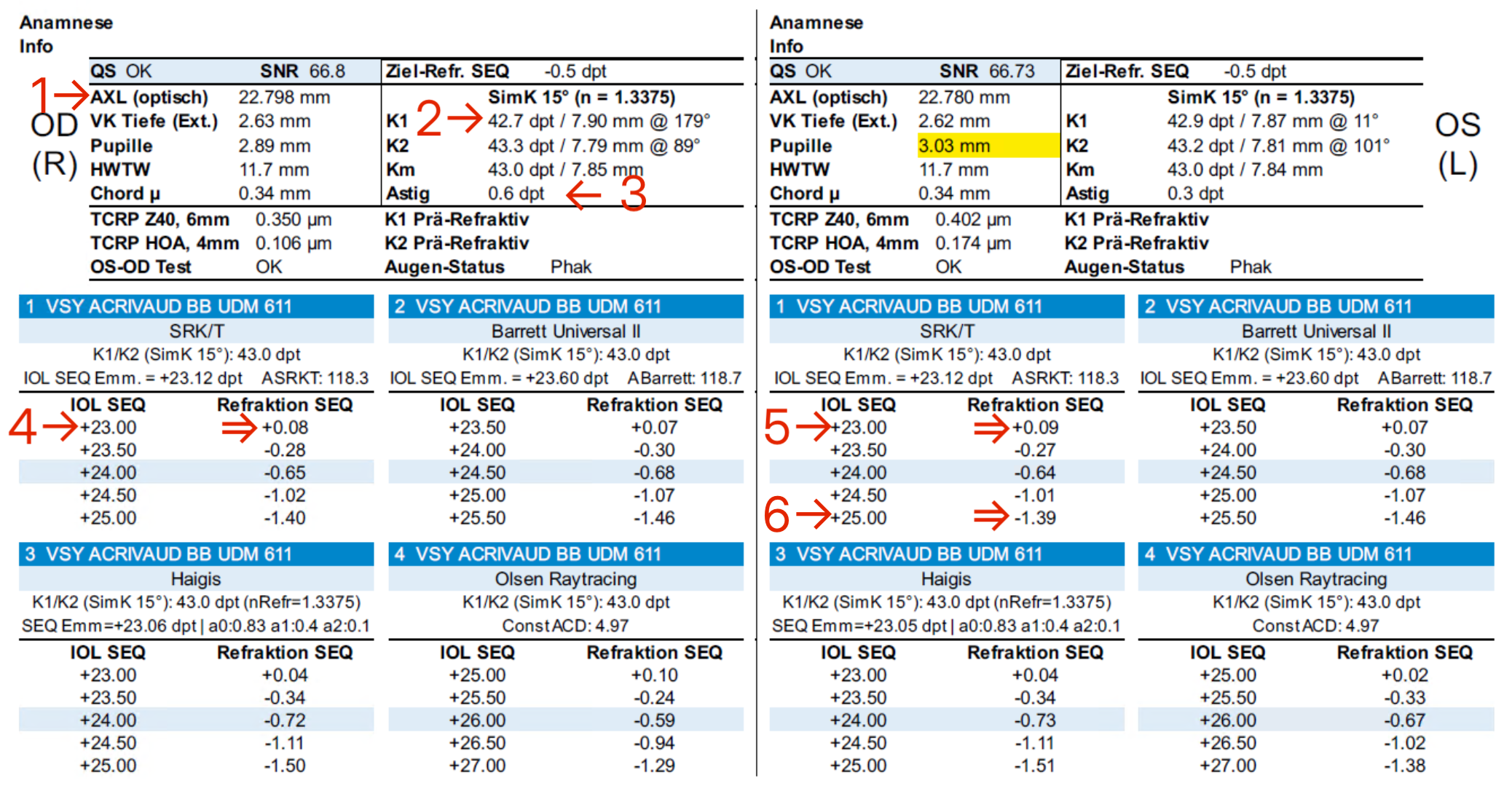
1. Normal axial length (22-24.5mm) 2. Normal K values (41.5-44 dioptres) 3. Astigmatism < 0.7 dioptres 4. Right eye: IOL for emmetropia +23.0 gives a refraction of +0.06 (closest to zero) 5. Left eye: +23.0 gives a refraction of +0.09 dioptres (closest to zero) 6. Left eye (non-dominant) for monovision: +25.0 gives a refraction of -1.39 dioptres (target refraction -1.25 to -1.5 dioptres)
Determining toric (tIOL) / multifocal IOL (mfIOL) power
Prerequisites
The patient wants maximum distance visual acuity without needing glasses, or wants monovision
Predominantly regular astigmatism with components at 90° (steep and flat axes at right angles to each other)
Stable topography
Keratoconus is usually stable at cataract age
In iatrogenic keratectasia or PMD, progression must generally be assumed -> no implantation of tIOLs or mfIOLs -> evaluate corneal cross-linking
Good visual potential (retina/optic nerve intact)
Contraindications
Severely irregular cornea (keratoconus, PMD, scar, keratectasia) -> implant a spherical/aspheric IOL, with touch-up by PRK and cross-linking if needed
Progressive keratectasia -> cross-linking
Strongly fluctuating topographies -> rule out progressive keratectasia
Status post keratoplasty (DALK/PKP)
Astigmatism should be corrected at the corneal level (re-suturing, opening the interface)
Add-on implantation/PIOL implantation in front of the spherical/aspheric IOL if needed
Progressive pterygium -> 1. pterygium excision 2. wait until topography is stable (at least two topographies after three weeks and after six weeks) 3. tIOL if appropriate
The patient has corneal astigmatism in both eyes and wants binocular distance vision, i.e. sharp distance vision with both eyes (ONO guideline: only available with the "Silver" or "Gold" package)
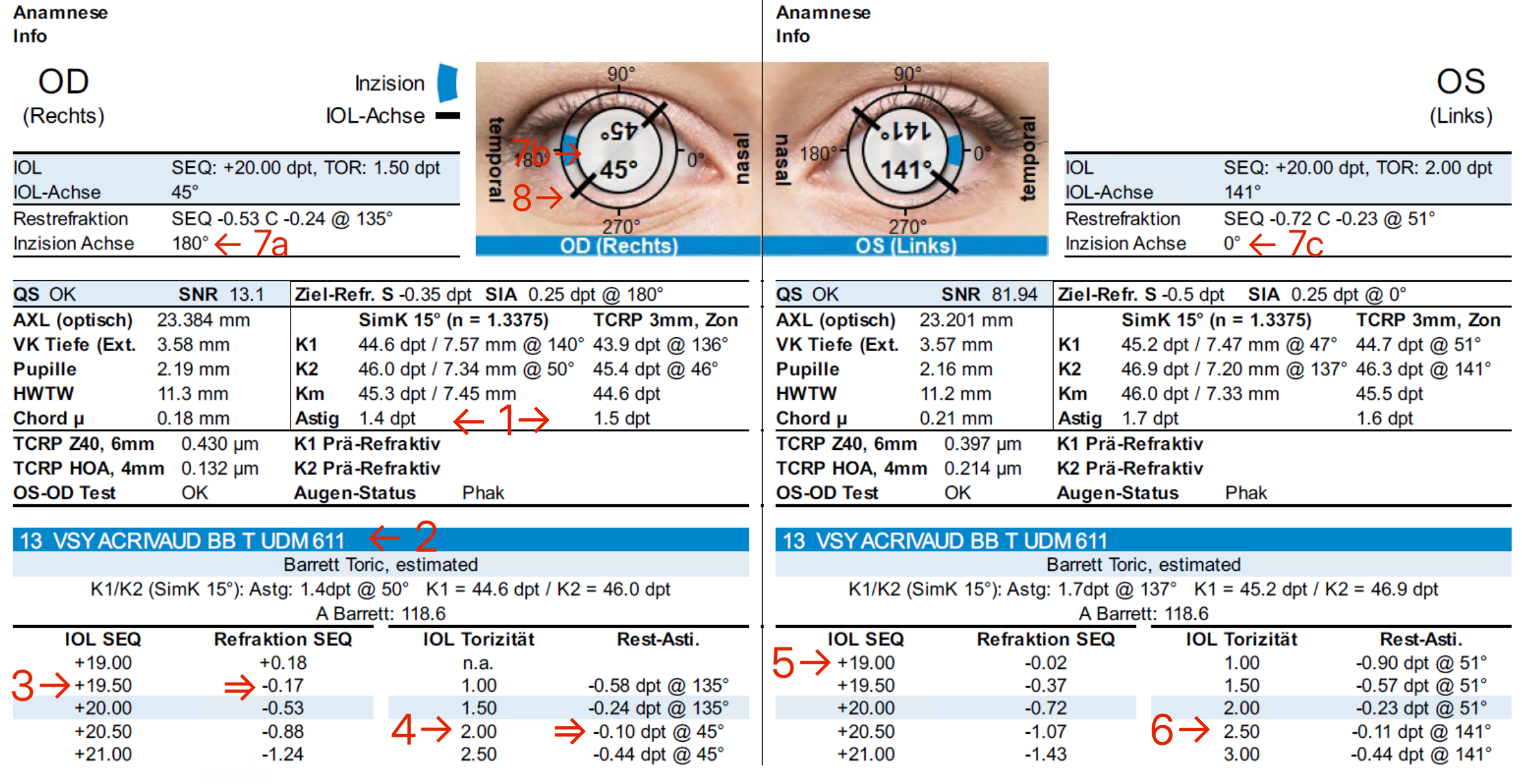
1. left (OD): anterior corneal astigmatism (SimK) 1.4 dioptres, to its right anterior astigmatism minus posterior astigmatism (Total Corneal Refractive Power, TCRP) > 0.7 dioptres -> toric IOL needed 2. UDM 611 BB Toric (has a different A-constant than the non-toric UDM 611) Right eye 3. Spherical IOL power: +19.5 gives a refraction of -0.17 (closest to zero) 4. IOL toricity of 2.0 gives a residual astigmatism of -0.1 (closest to zero) at 45°, i.e. the astigmatism is slightly overcorrected Left eye 5. Spherical IOL power: +19.0 gives -0.02 (closest to zero) 6. IOL toricity: 2.5 gives -0.11 (closest to zero) 7. Temporal incision 8. IOL implantation axis (aligned with the steep axis, since the IOL toricity is expressed as a plus cylinder)
Correct clinical note documentation
Template "vc"
Plan: Phaco-IOL by X Package: none/ Basic/ Vision/ Prime/ Deluxe Goal: binocular distance vision/ near vision/ monovision IOL: monofocal aspheric/ EDoF/ hybrid EDoF-MF 1. OX, ZR plan 2. OX, ZR plan Caution: monocle/ HPMC 2% on the table/ claustrophobia/ allergies ASA risk group: I/II/III no GP report required (ASA I or II and < 70 years old) Report to GP for premedication (ASA III or > 70 years old)
Template, worked example
Plan: Phaco-IOL by Dr. Muster in 4-6 weeks (e.g. earlier surgery in case of elevated intraocular pressure) Package: Vision (he does not want glasses for distance vision) Goal: binocular distance vision IOL: Monofocal Aspheric UDM 611 BB Toric (a toric IOL is required for astigmatism of > 0.7 dioptres) 1. OS plan, +19.5/2.5/141°vw (initials of the physician who determined the IOL) 2. OD, ZR plan +19.00/2.0/45°vw Caution: HPMC 2% on the table (due to reduced endothelial cell count)/ claustrophobia (needs more propofol)/ allergies: e.g. penicillin/iodine/latex ASA risk group: II (has arterial hypertension and mildly elevated blood glucose, both well controlled) No GP report required (< 70 years old) (is 69 years old)
Examples of IOL determination according to the ONO guideline
Example entry for the preoperative cataract assessment based on the "vc" template
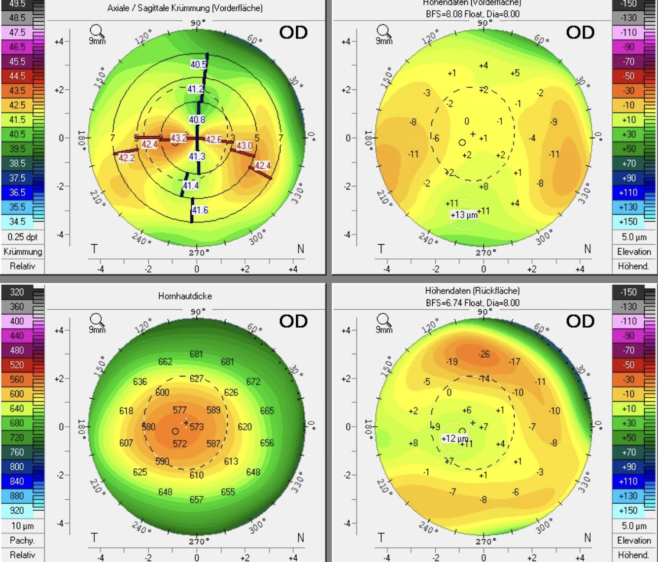
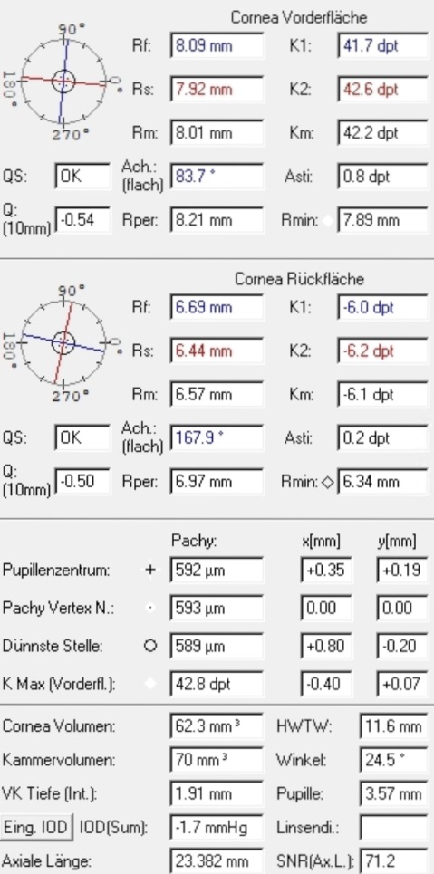
Corneal tomography using Pentacam® Right eye Assessment: relatively regular against-the-rule astigmatism
-> toric IOL indicated
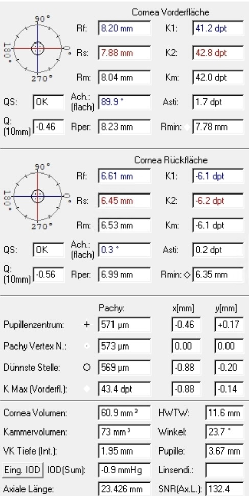
Values for IOL calculation
Left eye Assessment: regular against-the-rule astigmatism
-> toric IOL indicated
Topography data
IOL calculation with Pentacam ALX®
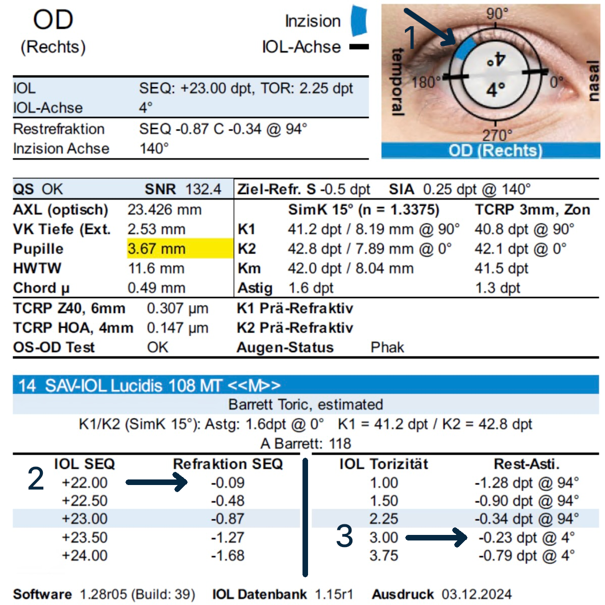
Example of a biometry with incision
Structured approach to IOL determination
1. Overview
Check that the incision is correct (temporal, superior, superotemporal)
Implantation axis = steep axis, i.e. plus cylinder (refraction is always expressed as minus cylinder -> flat axis)
2. Determining the spherical component of the IOL
as close to zero as possible (here +22.0 dioptres -> target refraction -0.09)
3. Determining toricity
IOL toricity of 1.0, 1.5, 2.25 dioptres -> astigmatism is undercorrected -> negative residual astigmatism remains on the refraction axis
as close to zero as possible, even if this results in a slight overcorrection (this is absolutely contraindicated when fitting glasses, but not with IOLs)
3.0 dioptres -> astigmatism is slightly overcorrected -> the axis of the residual astigmatism therefore rotates 90° to 4° (corresponding to the steep implantation axis)
the implantation axis is primarily determined by the steep axis (K2, example below: K2=0°) and may deviate from it slightly depending on the SIA (astigmatism induced by the incision) (see IOL axis=4°)
1. Overview sketch: superotemporal incision for a right-handed surgeon (light blue), implantation axis (here 4°) 2. Determining the sphere: +22.0 -> -0.09 (i.e. mild myopia) 3. Determining toricity
After corneal refractive surgery
Ablation of the cornea changes its curvature, i.e. the K values, which means that the usual formulas for IOL calculation are no longer accurate.
Several formulas take these changes into account. The ESCRS IOL calculation platform uses several formulas simultaneously, depending on which data are available. The more data available, the more accurate the calculation. Ideally, the patient's old K values and spectacle prescription should be obtained from them to optimise the calculation.
left: IOL calculation after LASIK/PRK for myopia middle: for hyperopia right: after radial keratotomy
Fill in the form for the IOL calculation -> Vertex distance 12 mm -> Device Keratometric Index (n) 1.3375
1. Physician and patient details 2. Pre-LASIK/PRK data (from old records) 3. Post-LASIK/PRK data (current measurements) -> for Pentacam® see below 4. Biometry data
Pentacam® -> From the topography -> corneal power distribution -> True Net Power at 4.0 mm
-> take from the biometry (important: internal AC depth, i.e. anterior chamber depth internal)
Special considerations
If, due to a previous refractive procedure, the patient has a decentred or small optical zone, it is advisable to aim the target refraction slightly towards hyperopia. A resulting hyperopic ablation profile is in this case better suited to regularising and enlarging the topography in a further step by means of excimer laser ablation (retreatment).